
Study compares antibody response in serum of patients who have recovered from COVID-19
Read Time:4 Minute, 31 Second
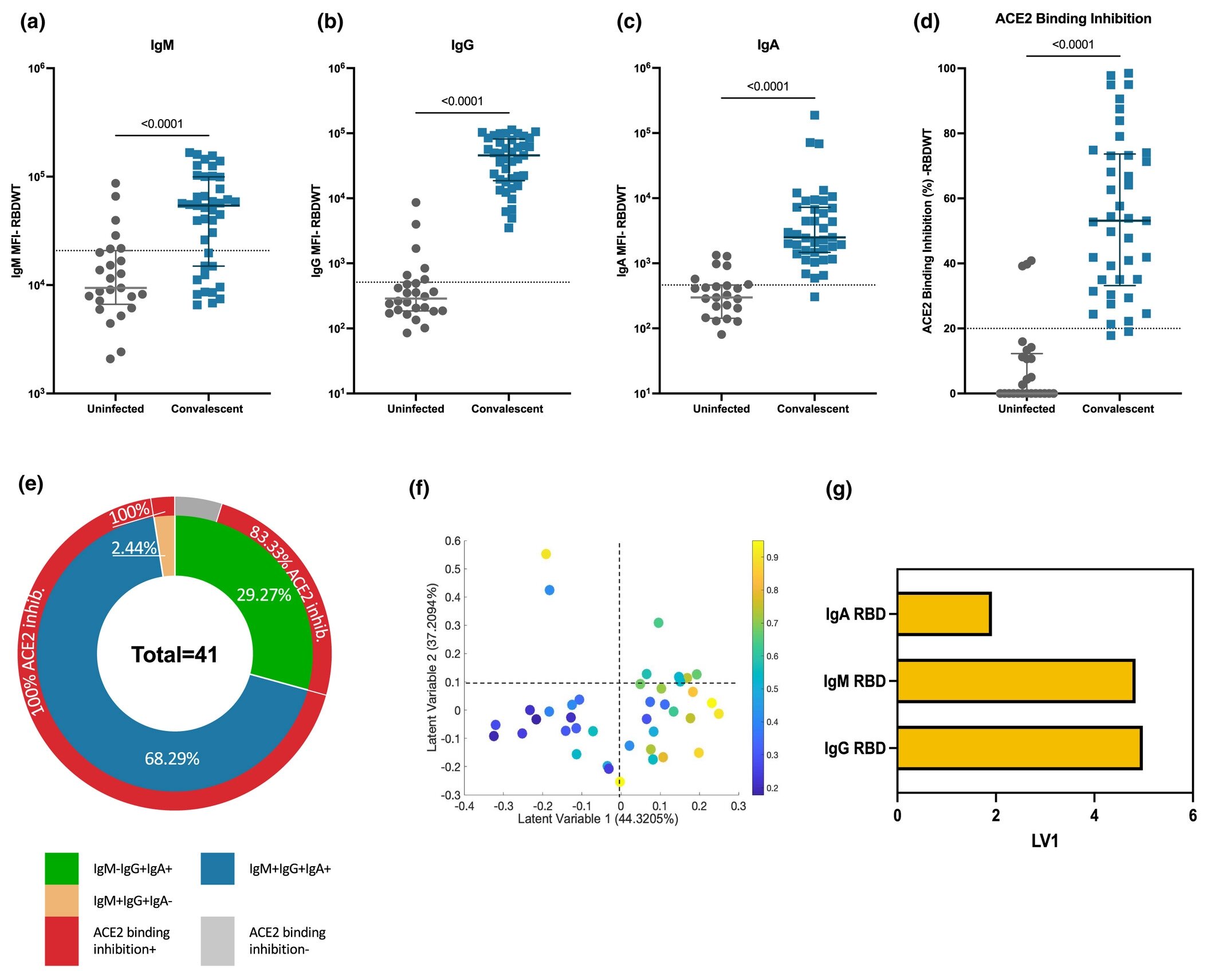
Convalescent plasma induced strong levels of anti-SARS-CoV-2 RBDWT antibody isotypes that interfere with ACE2 binding to RBDWT. Convalescent (n = 41) (blue) and uninfected control (n = 26) (gray) plasma (final dilution 1:100) was assessed for binding of IgM (a), IgG (b) and IgA (c) antibodies to SARS-CoV -2 RBDWT via multiplex. The positive threshold (gray dashed line) was defined as the 75th percentile of antibody binding (MFI) for uninfected control plasma. Statistical analysis was determined using the Mann-Whitney U-test. (d) RBDWT-ACE2 binding inhibition (%) of convalescent (blue) and uninfected control (grey) plasma (diluted 1:100). A positive threshold (gray dashed line) was defined as > 20% inhibition of ACE2 binding. (e) Pie chart showing percentage of subjects seropositive for anti-RBDWT antibody isotypes [IgM−IgG+IgA+ (green), IgM+IgG+IgA+ (blue), IgM+IgG+IgA− (yellow)] in the inner ring and the percentage of each seropositive subgroup with inhibition of ACE2 binding in the outer red ring. Partial least squares regression (PLSR) was performed to determine the multivariate relationship between anti-RBD-specific isotype antibodies (IgG, IgA, and IgM) and % inhibition of RBDWT-ACE2 binding (% inhibition of ACE2 binding is shown as a color gradient legend on the right yellow— the strongest to dark blue is the weakest). PLSR scores (f) and loadings plot (g). The percentage of variance for each latent variable (LV) is shown in parentheses. credit: Clinical and Translational Immunology (2022). DOI: 10.1002/cti2.1424
New research from the Peter Doherty Institute of Infection and Immunity (Doherty Institute), University of Melbourne and WEHI has revealed key information in our understanding of molecular immune responses to COVID-19.
After infection with COVID-19, virus-specific antibodies are generated that can both neutralize the virus and eliminate the infection. While much has been said about the importance of antibodies to immunoglobulin G (IgG) in protection and control of SARS-CoV-2, the role of antibodies to immunoglobulin A (IgA) against SARS-CoV-2 has been relatively neglected throughout the pandemic. Until now.
In the article published on October 23 Clinical and Translational Immunologyscientists conducted an experimental study to compare the response of antibodies to the virus blood serum from people who have recovered from COVID-19.
Samantha Davies of the University of Melbourne, Ph.D. Researcher at the Doherty Institute, lead author of the paper.
“Simply put, we deconstructed blood in our lab to measure its ability to suppress the virus and activate immune cells to destroy SARS-CoV-2″- explained Davis.
“While we knew that IgG was very important in the antibody response to clear the virus, we found that IgA also plays a key role in neutralizing it in most people.”
As neutralizing antibodies are an indicator of immune response and protection against viral infectionsthis finding is critical in the context of vaccine development.
Dr Amy Chung of the University of Melbourne is the head of the Doherty Institute laboratory and one of the senior authors of the paper.
“This study opens the door to new approaches for the development of future vaccines against SARS-CoV-2″Dr. Chang said.
“Our findings are particularly important because IgA is the most common antibody present in the lining of our respiratory tract, which is the main route for the virus to spread. infection. This means that if we can specifically produce these antibodies at these vulnerable sites, we now know that we can induce a robust immune response to protect against virus.”
Additional information:
Samantha K. Davis et al. Heterologous reactions of antibodies neutralizing SARS-CoV-2 IgA in convalescent plasma, Clinical and Translational Immunology (2022). DOI: 10.1002/cti2.1424
Samantha K. Davis et al. Heterologous reactions of antibodies neutralizing SARS-CoV-2 IgA in convalescent plasma, Clinical and Translational Immunology (2022). DOI: 10.1002/cti2.1424
Courtesy of the Peter Doherty Institute of Infection and Immunity
Citation: Study compares serum antibody responses of patients who have recovered from COVID-19 (Oct. 25, 2022) Retrieved Oct. 25, 2022, from https://medicalxpress.com/news/2022-10-antibody-responses-blood -serum-patients. html
This document is subject to copyright. Except in good faith for the purpose of private study or research, no part may be reproduced without written permission. The content is provided for informational purposes only.
Happy
0 %
Sad
0 %
Excited
0 %
Sleepy
0 %
Angry
0 %
Surprise
0 %